
Bone grafting is often closely associated with dental restorations such as bridge work and dental implants. In the majority of cases, the success of a restoration procedure can hinge on the height, depth, and width of the jawbone at the implant site. When the jawbone has receded or sustained significant damage, the implant(s) cannot be supported on this unstable foundation and bone grafting is usually recommended for the ensuing restoration.
Over a period of time, the jawbone associated with missing teeth atrophies or is reabsorbed. This often leaves a condition in which there is poor quality and quantity of bone suitable for placement of dental implants. In these situations, most patients are not candidates for placement of dental implants. Today, we have the ability to grow bone where needed. This not only gives us the opportunity to place implants of proper length and width, it also gives us a chance to restore functionality and aesthetic appearance.
Periodontal Disease – Periodontal disease can affect and permanently damage the jaw bone that supports the teeth. Affected areas progressively worsen until the teeth become unstable.
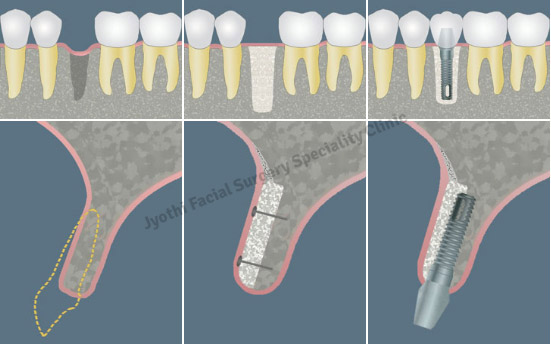
Tooth Extraction – Studies have shown that patients who have experienced a tooth extraction subsequently lose 40-60% of the bone surrounding the extraction site during the following three years. Loss of bone results in what is called a “bone defect”.
Injuries and Infections – Dental injuries and other physical injuries resulting from a blow to the jaw can cause the bone to recede. Infections can also cause the jaw bone to recede in a similar way.
Bone grafting is a highly successful procedure in most cases. It is also a preferable alternative to having missing teeth, diseased teeth, or tooth deformities. Bone grafting can increase the height or width of the jawbone and fill in voids and defects in the bone. There are essentially two basic ways in which bone grafting can positively impact the health and stability of the teeth:
Jaw Stabilization – Bone grafting stabilizes and helps restore the jaw foundation for restorative or implant surgery. Deformities can also be corrected and the restructuring of the bone can provide added support.
Preservation – Bone grafting can be used to limit or prevent bone recession following a tooth extraction, periodontal disease, or other invasive processes.Initially, the surgeon will thoroughly examine the affected area in order to assess the general condition of the teeth and gums. If periodontal disease is present or the adjacent teeth are in poor condition, these factors will be fully addressed before the bone grafting procedure can begin. The surgeon will also recommend panoramic x-rays in order to assess the precise depth and width of the existing bone. On occasion, a CT scan may be recommended to determine the bone condition. Depending on these results, the surgeon may also anesthetize the area and explore into the gum in order to determine what kind and how much bone is required.
There are several types of bone grafts. Your surgeon will determine the best type for your particular condition.
Autogenous Bone Graft - Harvested from the patient’s own body (usually from the posterior part of the lower jaw or the chin). This method is usually preferred because it produces the most predictable results.
Allogenic Bone Graft - Cadaver or synthetic bone is used in this type of graft.
Xenograft - Cow bone is used in this type of graft.
The bone grafting procedure can often take several months to complete. Bone is typically harvested from your own body and added to the affected site. This bone will fuse with the existing bone and the migration of cells will cause firm adhesion and cell growth. Supplementing the jaw with bone will result in greater bone mass to help support and anchor the implant(s).
During the surgery, the surgeon will numb the grafting and extraction sites using local anesthetic. A small incision will be made to prepare the site for the new bone and it will be anchored into place. On occasion, a synthetic membrane may be used to cover the new bone. This membrane prevents soft tissue and bacterial invasions, and encourages new bone growth. The surgery does not require an overnight stay, and you will be provided with comprehensive instructions for your post-operative care. The Surgeon will prescribe medications to help manage infection, discomfort and swelling.
Bone grafting can repair implant sites with inadequate bone structure due to previous extractions, gum disease or injuries. The bone is either obtained from a tissue bank or your own bone is taken from the jaw, hip or tibia (below the knee). Sinus bone grafts are also performed to replace bone in the posterior upper jaw. In addition, special membranes may be utilized that dissolve under the gum and protect the bone graft and encourage bone regeneration. This is called guided bone regeneration or guided tissue regeneration.
Major bone grafts are typically performed to repair defects of the jaws. These defects may arise as a result of traumatic injuries, tumor surgery, or congenital defects. Large defects are repaired using the patient’s own bone. This bone is harvested from a number of different sites depending on the size of the defect. The skull (cranium), hip (iliac crest), and lateral knee (tibia), are common donor sites. These procedures are routinely performed in an operating room and require a hospital stay.
The maxillary sinuses are behind your cheeks and on top of the upper teeth. Sinuses are like empty rooms that have nothing in them. Some of the roots of the natural upper teeth extend up into the maxillary sinuses. When these upper teeth are removed, there is often just a thin wall of bone separating the maxillary sinus and the mouth. Dental implants need bone to hold them in place. When the sinus wall is very thin, it is impossible to place dental implants in this bone. There is a solution and it’s called a sinus graft or sinus lift graft. The dental implant surgeon enters the sinus from where the upper teeth used to be. The sinus membrane is then lifted upward and donor bone is inserted into the floor of the sinus. Keep in mind that the floor of the sinus is the roof of the upper jaw. After several months of healing, the bone becomes part of the patient’s jaw and dental implants can be inserted and stabilized in this new sinus bone.
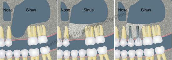
The sinus graft makes it possible for many patients to have dental implants when years ago there was no other option other than wearing loose dentures. If enough bone between the upper jaw ridge and the bottom of the sinus is available to stabilize the implant well, sinus augmentations and implant placement can sometimes be performed as a single procedure. If not enough bone is available, the sinus augmentation will have to be performed first, then the graft will have to mature for several months, depending upon the type of graft material used. Once the graft has matured, the implants can be placed.
In severe cases, the ridge has been reabsorbed and a bone graft is placed to increase ridge height and/or width. This is a technique used to restore the lost bone dimension when the jaw ridge gets too thin to place conventional implants. In this procedure, the bony ridge of the jaw is literally expanded by mechanical means. Bone graft material can be placed and matured for a few months before placing the implant.
The inferior alveolar nerve, which gives feeling to the lower lip and chin, may need to be moved in order to make room for placement of dental implants in the lower jaw. This procedure is limited to the lower jaw and indicated when teeth are missing in the area of the two back molars and/or and second premolar, with the above-mentioned secondary condition. Since this procedure is considered a very aggressive approach (there is almost always some postoperative numbness of the lower lip and jaw area, which dissipates only very slowly, if ever), usually other, less aggressive options are considered first (placement of blade implants, etc.).
Typically, an outer section of the cheek side of the lower jawbone is removed in order to expose the nerve and vessel canal. Then we isolate the nerve and vessel bundle in that area and slightly pull it out to the side. At the same time, we will place the implants. Then the bundle is released and placed back over the implants. The surgical access is refilled with bone graft material of the surgeon’s choice and the area is closed.
These procedures may be performed separately or together, depending upon the individual's condition. As stated earlier, there are several areas of the body that are suitable for attaining bone grafts. In the maxillofacial region, bone grafts can be taken from inside the mouth, in the area of the chin or third molar region, or in the upper jaw behind the last tooth. In more extensive situations, a greater quantity of bone can be attained from the hip or the outer aspect of the tibia at the knee. When we use the patient’s own bone for repairs, we generally get the best results.
In many cases, we can use allograft material to implement bone grafting for dental implants. This bone is prepared from cadavers and used to promote the patients own bone to grow into the repair site. It is quite effective and very safe. Synthetic materials can also be used to stimulate bone formation. We even use factors from your own blood to accelerate and promote bone formation in graft areas. These surgeries are performed in the out-office surgical suite under IV sedation or general anesthesia. After discharge, bed rest is recommended for one day and limited physical activity for one week.